


Chigbogu P. Nwankwocha, Elijah S. Okwuonu, Chinaza B. Ukwueze, Adaobi O. Opah, Ifeanyi D. Ogbonna, Chioma P. Ezeme & Patience O. Ubachukwu
Abstract
Background
Malaria remains a significant public health concern, disproportionately impacting vulnerable populations in resource-limited settings.
Methods
A study conducted in Nkanu West Local Government Area examined malaria prevalence and related anemia among children aged 6–10 years and among pregnant women attending a hospital. The research employed a cross-sectional, hospital-based design, using both microscopy and rapid diagnostic tests (RDTs) to evaluate malaria cases. Microscopy enabled detailed detection of parasites, while RDTs provided quick and supplementary diagnostics.
Result
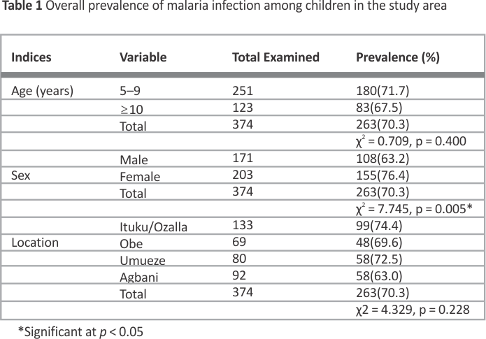
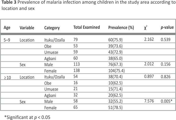
This study revealed an alarming malaria prevalence of 70.3% in children and 65.5% in pregnant women. Microscopy prevalence slightly increased with age (r = 0.118, p < 0.05), while RDT decreased (r=-0.157, p < 0.01). Among 10-year-olds and above, females had a significantly higher prevalence (78.5%) than males (55.2%) (χ²=7.576, p < 0.05). There were significant differences in prevalence by sex (p = 0.005), but not by age (p = 0.400) and location (p = 0.228) among children. Among pregnant women, no significant differences were found concerning age, trimester, or location. Malaria was linked to increased packed cell volume, especially in women in their third trimester, indicating hematological impact. Mean PCV of children with malaria varies significantly by age, with 5–9-year-olds having a higher mean PCV (37.65 ± 0.90%) than other age groups (t = 2.434, p = 0.059).
Conclusion
Malaria remains a threat to children and pregnant women, requiring ongoing monitoring, community awareness, routine testing, and sanitation, especially during ante- natal care. Based on our findings, priority interventions should include universal ITN distribution with sustained use, intensified vector control and larval source management in high-burden areas, strengthened IPTp delivery and ANC-based screening, environmental sanitation and waste management to curb breeding sites, and enhanced community surveillance to guide adaptive program planning.
1 Background
Malaria remains a significant public health challenge in Nigeria, primarily transmitted by female Anopheles mosquitoes infected with Plasmodium species. Among these, P. falciparum accounts for approximately 98–99% of cases (WHO, 2022). Other species, such as P. malariae and P. ovale, also contribute to the disease burden. The principal mosquito vector species present in Nigeria, Anopheles gambiae, Anopheles funestus, Anopheles arabiensis, and Anopheles melas, are distributed throughout the country’s primary ecological zones, which encompass Mangrove, Freshwater Swamp, Rain Forest, Derived Savannah, Guinea Savannah, Sudan Savannah, Sahel Savannah, and Mid-Altitude regions. An. gambiae and An. funestus exhibit widespread distribution, whereas An. arabiensis is typically found in drier environments, and An. melas is predominantly associated with coastal mangrove ecosystems. The impact of malaria is extensive, responsible for 30–40% of outpatient visits, around 20% of childhood deaths, and significant maternal mortality, which exerts immense pressure on Nigeria’s healthcare system 4. Socioeconomic effects include school absenteeism, reduced productivity, and an estimated annual cost of 132 billion Naira 20. Despite global progress, persistent malaria transmission in Nigeria underscores the urgent need for intensified control strategies aligned with the Sustainable Development Goals, targeting malaria elimination by 2030 (UN, 47; 14. While there have been studies on malaria prevalence and anemia in Nigeria, few have focused on rural settings like Nkanu West, where malaria transmission is high, and access to healthcare services is limited.
The pathophysiology of malaria is complex, especially regarding its most severe complication, malaria anemia. Parasites induce hemolysis by replicating inside and rupturing infected red blood cells (RBCs). Additional mechanisms include dysregulation of erythropoiesis, destruction of non-parasitized RBCs (nRBCs), increased apoptosis, and senescence of RBCs 37. The immune response further accelerates RBC destruction through processes such as opsonization and complement activation. Nutritional deficiencies, particularly of iron, folate, and vitamin B12, exacerbate anemia, impairing recovery and elevating morbidity 19,28. High parasite loads often impair erythropoiesis in the bone marrow, contributing to severe anemia, which is particularly prevalent in areas with high transmission rates 29. Vulnerable groups include young children and pregnant women, with severe cases often requiring blood transfusions during the rainy season when transmission peaks 24.
Clinical manifestation of malaria varies across Nigeria. For example, cerebral malaria sequelae are more common in the north, whereas severe anemia due to malaria predominates in the south 22,41. Malaria transmission remains intense and stable nationwide, with classification based on spleen rates in children: hypoendemic (< 10%), mesoendemic (11–50%), hyperendemic (> 75% in children and > 25% in adults), and holoendemic (> 75% in children with low adult rates). Rural areas often experience holoendemic transmission with persistent high rates, while urban centers tend to be mesoendemic (WHO, 53). Seasonality of malaria influences transmission intensity, incidence during the rainy season in the north and year-round in the south 54.
Control measures aimed to reduce morbidity and mortality but face challenges in measuring their true impact across sub-Saharan Africa (WHO, 2023). Parasite density correlates with transmission intensity, with higher densities found in areas of high endemicity 23. Malaria often results in anemia, characterized by reduction in hemoglobin or RBC count below normal levels (WHO, 2022). Anemia in malaria patients is typically normocytic and normochromic, but can also be microcytic and hypochromic due to underlying hemoglobinopathies and iron deficiency prevalent in endemic regions 7, 26.
Clinically, anemia may manifest as fatigue, shortness of breath, pallor, and palpitations; severe anemia (hemoglobin < 70 g/L in children under five; <80 g/L in older individuals) is life-threatening and can lead to heart failure or death 48. Diagnosis primarily involves measuring hemoglobin levels, which vary according to age, sex, pregnancy status, environmental factors (e.g., altitude and smoking), and genetics. WHO’s hemoglobin cut-offs for anemia are globally adopted but adjusted to reflect demographic and physiological differences. For populations of African descent, base line hemoglobin levels tend to be lower, partly due to genetic hemoglobin disorders like sickle cell anemia, requiring tailored thresholds 2.
In malaria-endemic areas, more than half of malaria-related deaths can be attributed to severe anemia. WHO recommends using anemia prevalence as an indirect marker for malaria burden at the community level due to its sensitivity to changes in transmission (WHO, 2025). Children are especially vulnerable, with anemia significantly contributing to hospital admissions during peak seasons 15. Pregnant women face increased risks due to immunological modifications and placental sequestration of parasites, which raise their chances of severe anemia and adverse birth outcomes like low birth.
Understanding the prevalence and interplay of malaria and anemia in this setting is vital for designing effective control strategies and monitoring their impact over time. Given that malaria-related severe anemia accounts for significant proportions of childhood morbidity and mortality, especially in endemic regions like Nigeria, this study contributes to refining the current indicators used for malaria burden assessment by providing data-driven insights, which relate to Nigeria’s goal of reducing malaria burden and achieving elimination by 2030. Despite ongoing malaria control programs in Nigeria, Nkanu West LGA experiences persistently high malaria prevalence, particularly among children and pregnant women. This study seeks to explore the region-specific factors contributing to this trend.
2 Methods
2.1 Study area
The study was conducted in Nkanu West LGA, Enugu State, Nigeria, with headquarters in Agbani. Covering 225 km² in the tropical savanna zone, it has a population of about 211,500 (NPC, 34). Temperatures range from (23.1–31) °C, with annual rainfall of 1520–2030 mm. The region has a rainy season (March – October) and a dry season (November– February). Most residents are farmers cultivating yams, cassava, maize, rice, and vegetables, living in dispersed compounds surrounded by farmland and trees, e.gs, palm, mango, and banana, which increases exposure to mosquito bites and malaria risk.
2.2 Study design
This descriptive observational study was carried out at the University of Nigeria Teaching Hospital, Ituku/Ozalla. Ethical clearance was obtained from the institutional-based review committee. The research team, including researchers, phlebotomists, and lab scientists, collected blood samples after obtaining consent from pregnant women and caregivers of children. Participants were randomly selected from the hospital’s outpatient Hematology Clinic, ensuring balanced representation of children and pregnant women from different socioeconomic backgrounds and geographical locations within the LGA.
2.3 Study population and sample size
The minimum sample size was calculated to be 487 participants, comprising 374 children and 113 pregnant women, based on standard prevalence and statistical formulas with a 95% confidence level and 5% margin of error 5. Therefore, 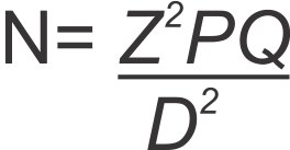
where Z = 95% (1.96), P = 58% (0.58), q = 1- 0.58 (0.42), d = 5% (0.05); N=374
2.4 Procedure for sample collection
A total volume of 2 ml of venous blood was drawn from each participant using standard techniques. A tourniquet was tied around the upper arm after cleaning the site with an antiseptic wipe. A sterile needle was inserted into the vein, and blood was collected into EDTA anticoagulant bottles. After collection, the tourniquet was released, and pressure was applied with cotton wool to stop bleeding, followed by a plaster to secure the site. EDTA prevents blood clotting by chelating calcium, ensuring suitability for hematological analysis.
2.5 Preparation of blood films
Both thick and thin blood films were prepared for analysis.
2.6 Procedure for thin film Preparation for malaria parasite examination
A small volume of anti-coagulated blood was placed about 2 cm from the slide edge. A spreader, held at a 45° angle, was gently applied to the blood droplet, spreading it evenly. The film was then fixed with methanol after air-drying. The slide was stained with alpha-phenolphthalein stain to preserve parasite morphology, aiding identification 10. After staining, the slide was rinsed with clean water to remove excess stain for microscopic examination.
2.7 Microscopy examination for malaria parasites
A high-power microscope, typically magnified between 400 and 1,000 times using an oil immersion objective, is employed to analyze both thick and thin blood films 9, 44,45 WHO, 2010). This technique remains the gold standard for detecting and identifying malaria parasites in laboratory settings.
2.8 Rapid diagnostic test
A Malaria Plasmodium antigen detection kit (manufactured by SD Bioline, India) was utilized for diagnostic purposes. The kit includes a test cassette, buffer, and pipette. Using the pipette, two drops of blood from the EDTA tube were placed on the cassette, followed by three drops of buffer. After 15 minutes of incubation, the results were read: two lines (control and test) indicated a positive malaria antigen, while a single control line indicated a negative result. While microscopy offers higher sensitivity and specificity, RDTs provide a quick and efficient screening method, making them complementary tools in this study for faster diagnosis and ensuring accuracy in a hospital setting.
2.9 Parasite density assessment
This was determined from Giemsa-stained thick blood smears by enumerating asexual parasites against 200 leukocytes and expressing the result as parasites per microliter of blood (p/µL) 9. An assumed leukocyte concentration of 8,000/µL was applied to convert counts to density, using the formula: Parasite density (p/µL) = (parasites counted/200) × 8,000. An independent second reader re-evaluated 10% of the slides to assess reliability, with any discrepancies resolved by a third reader. Slides exhibiting poor staining or unreadable fields were excluded from density analyses, and reported densities pertain only to slides of adequate quality.
2.10 Packed cell volume
Hematocrit was used to screen for anemia, suitable for large clinic populations 10. The measurement involved centrifuging well-mixed, anti-coagulated blood in a capillary tube to separate RBCs. The tube was filled about three-quarters, sealed with plasticine, and placed in a centrifuge with the sealed end away from the center. The centrifuge was spun for 5 minutes, then stopped, and the PCV was read using a hematocrit tube reader. The percentage of blood volume occupied by RBCs ws calculated, providing the PCV value for each child and pregnant woman.

Data were analyzed with SPSS version 23.0. Malaria prevalence
was assessed using Chi-Square (IBM Corporation, Armonk, USA). Hematological differences were evaluated with ANOVA and Duncan’s New Multiple Range Test. The Student’s t-test compared PCV between sexes, while Pearson correlation examined associations between age, parasite infection, and demographic variables.
3 Results
3.1 Prevalence of malaria parasite infection among children and pregnant women
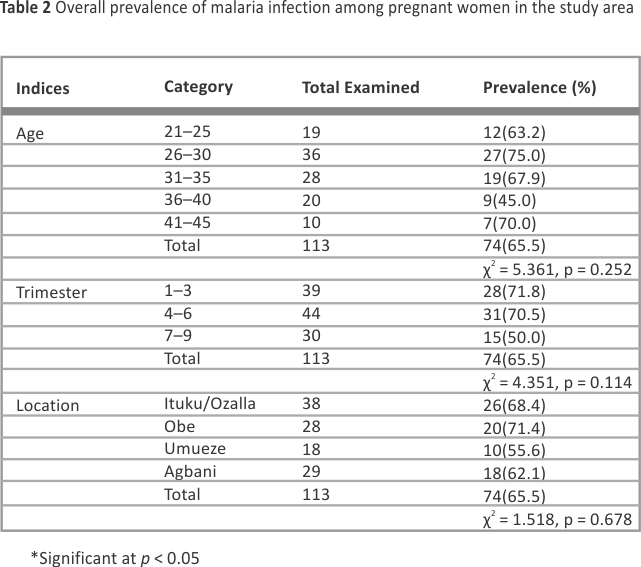
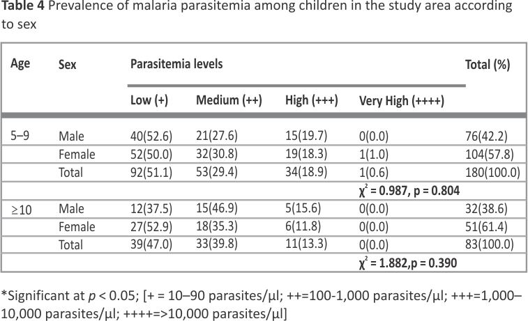
Overall, malaria prevalence was 70.3% in children and 65.5% in pregnant women. Significant differences in prevalence were observed by sex (p = 0.005), but not by age (p = 0.400) and location (p = 0.228) among children (Table 1). Among pregnant women, no significant differences were found concerning age, trimester, or location. However, higher infection rates were noted in women aged 26–30 years (75.0%), in the first trimester (71.8%), and residing in the Obe community (71.4%) (Table 2).
There were no significant differences (p > 0.05) in malaria prevalence among pregnant women across locations or trimesters (Table 5). Notably, higher prevalence was observed among first-trimester women in Ituku/Ozalla (85.7%) and Umueze (80.0%), while Agbani had a higher prevalence in the second trimester (70%). Malaria parasitemia levels did not significantly vary by age or pregnancy stage. Low parasitemia was more common in Ituku/Ozalla (42.3%), Obe (40.0%), and Umueze (50.0%) (Table 6).
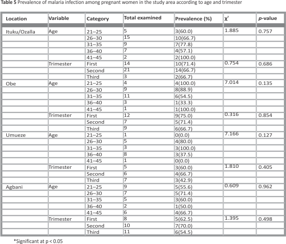
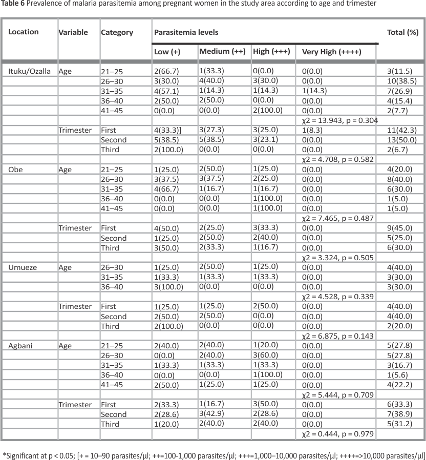
Figure 1 shows that the mean PCV of children with malaria varies significantly by age, with 5–9-year-olds having a higher mean PCV (37.65 ± 0.90%) than other age groups (t = 2.434, p = 0.059). No significant difference in PCV was observed between boys and girls (t = 0.267, p = 0.790). Figure 2 shows that among children with malaria within ages of 5–9 years, there was no significant difference in PCV levels between males and females (t = 0.913, p = 0.397). Conversely, male children who were 10 years old and above had higher PCV (35.84 ± 0.90%) than females (33.22 ± 0.52%) (t = 2.720, p = 0.008).
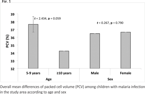
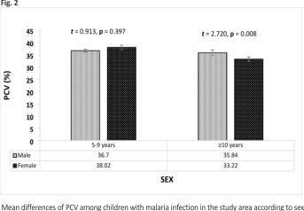
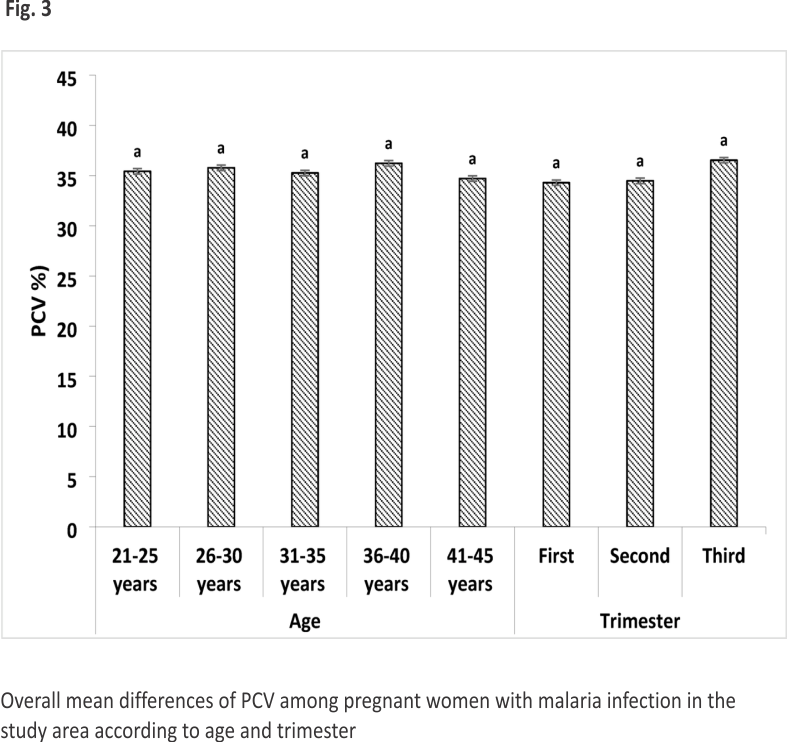
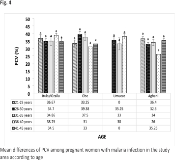
Table 7 shows that malaria parasitemia weakly and inversely correlates with children’s PCV (r–0.391, p < 0.01), and age also weakly negatively correlates with PCV (r=–0.203, p < 0.01). Table 8 indicates that among pregnant women, malaria parasitemia strongly and negatively correlates with PCV (r =–0.767, p < 0.01).
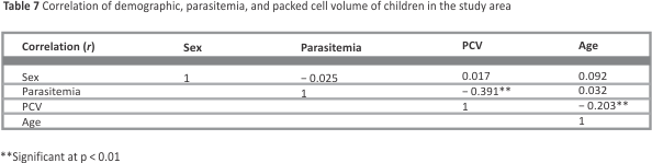
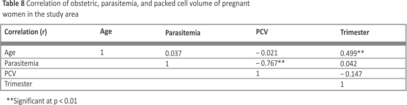
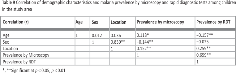
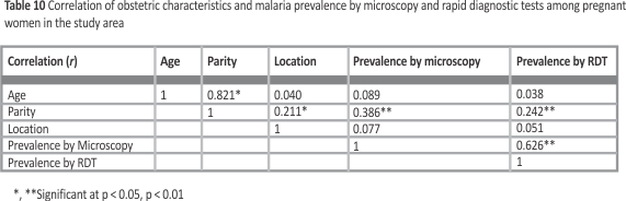
Table 9 shows that age moderately correlates with malaria prevalence (p < 0.01). Malaria prevalence by microscopy positively correlates with age (r = 0.118, p < 0.05), while RDT prevalence inversely correlates (r=−0.157, p < 0.01). Microscopy sensitivity increased with age, whereas RDT was more sensitive among younger children. There is a strong positive correlation between microscopy and RDT (r = 0.659, p < 0.01). Table 10 indicates that pregnancy trimester moderately correlates with malaria prevalence (p < 0.01). Detection by microscopy (r = 0.386) and RDT (r = 0.242) both significantly correlate with trimester (p < 0.01). The correlation between microscopy and RDT is also strong (r = 0.626, p < 0.01).
This hospital-based survey examined malaria-related anemia among children aged 6–10 years and pregnant women in Nkanu West LGA, Nigeria. The overall malaria prevalence was 70.3% among children, closely aligning with 71.1% reported by 6 in semi-urban communities in Southwestern Nigeria. However, the prevalence was higher than reported in some other Nigerian studies in the hospital environment 38. These findings reinforce the World Health Organization and Nigeria Malaria Indicator Survey’s assertion that malaria remains a significant public health issue globally, especially among children and pregnant women, necessitating integrated control strategies and broader efforts toward universal health coverage (NMEP, 33; WHO, 49).
Studies from Ogun, south western Nigeria, and South Sudan reported higher prevalence rates of 80.5% and 78%, respectively 40,46. Variations in malaria prevalence within Nigeria, from less than 20% in some regions to over 70% in others 43, are influenced by geographical differences, socio-economic challenges, and disparities in malaria control interventions. The elevated prevalence among children in this study may reflect environmental factors, like proximity to water bodies, drainage issues, refuse sites, and water-retaining containers, which favor mosquito breeding, especially during the rainy season.
Among pregnant women, the overall malaria prevalence was 65%. Higher rates have been reported in other Nigerian studies; for example, in Port Harcourt, 13 recorded 72%, Benin City 78.9% 39, while Kano State and Ile-Ife had lower figures of 39.2% 17 and 13.1% 16. Most research focused on women attending antenatal clinics, where routine screening and prophylaxis are common, highlighting the importance of healthcare access in malaria control among pregnant women. 25.
Our findings align with high-transmission figures in different six geopolitical zones in Nigeria 11, 12,18,42 MIS northcentral, northwest, & southeast, 2021; 27,41, though pronounced regional variability persists across the country. Environmental factors—such as proximity to water bodies, drainage challenges, and waste accumulation—likely sustain vector habitats. Heterogeneous implementation of preventive measures (ITN ownership and use, IPTp uptake, and ANC-based screening) and disparities in health service access likely drive observed regional differences between children and pregnant women prevalence. To reduce regional disparities and move toward universal health coverage, integrated malaria control is needed like sustained vector-control and environmental management, universal ITN distribution with reliable usage, and strengthening IPTp delivery through ANC services, complemented by triangulated surveillance that combines hospital-based data with community prevalence and intervention coverage indicators.
Location-specific analysis showed that Ituku/Ozalla had the highest malaria prevalence in children (74.4%), followed by Obe community in pregnant women (71.4%). These high rates likely result from environmental conditions conducive to mosquito breeding, such as water-retaining containers, bushes, drainage areas, and waste sites in these communities. Proximity to breeding habitats influences transmission rates, with environmental factors e.g., temperature and humidity also playing roles.
Moreover, the highest prevalence among pregnant women was observed in the 26–30-year age group (75.0%), consistent with 3 finding, who reported similar trends in rural Nigeria. Although no significant differences in malaria prevalence among pregnant women were observed concerning age and trimester, these factors influence anemia and hemorrhagic risks. Notably, the study found that PCV, an indicator of anemia, was non-significantly higher among women in their third trimester (38.00 ± 0.58%). This aligns with 25 work, indicating that anemia prevalence peaks during early pregnancy stages, especially in holoendemic regions, due to increased parasite density and immune suppression. Behavioral and socioeconomic factors significantly impact malaria risk. Socioeconomic status influences access to prevention tools, healthcare services, and information; higher socioeconomic groups typically have better access to protection and housing, thus reducing vulnerability. Poorer communities often lack proper infrastructure, waste management, and preventive resources, leading to higher mosquito densities and increased transmission risks 21.
However, public health interventions aimed at reducing mosquito breeding and expanding ITN usage in rural communities e.gs., Ituku/Ozalla, Obe, and nearby communities could markedly cut local malaria transmission and prevalence. Adopting an integrated vector management approach— encompassing environmental sanitation and universal ITN coverage—is likely to lower malaria prevalence in these high-burden pockets. Enhancing community-led vector control and guaranteeing ongoing ITN access in affected areas may produce meaningful declines in malaria burden across Nkanu West LGA. Findings from comparable settings support these strategies, with ITN scale-up linked to reduced malaria burden in northern Nigeria 1.
Furthermore, the Enugu State Government is expanding free, quality malaria services for children and pregnant women. By deploying 549 facilities with antimalarial drugs and testing, vulnerable households gain free diagnosis and treatment, reducing out-of-pocket costs and disease burden. This subsidized access, alongside bed nets, demonstrates a strong commitment to maternal and child health (NAN, 2025; 36.
The study’s findings on age-related differences in PCV values among children with malaria are noteworthy. Children aged 5–9 years had higher PCV values compared to older children, likely due to the development of partial immunity and reduced anemia severity. This observation highlights the importance of tailored diagnosis, treatment, and management approaches for different age groups ( Kokori et al., 2025).
The analysis of PCV levels among children showed no significant difference based on sex, consistent with previous studies 15. Malaria causes anemia through various mechanisms, including hemolysis of infected and uninfected RBCs, shortened RBC lifespan, and impaired RBCs in the bone marrow (CDC, 8). Chronic malaria infections often lead to dyserythropoiesis, further contributing to anemia prevalence.
The findings of this study have implications for malaria control and prevention strategies in the region. The use of insecticide-treated nets, proper waste management, and access to healthcare services are critical in reducing malaria transmission and its complications. The findings also underscore the need for location-specific analysis and targeted approaches to address the unique challenges and risk factors in different communities.
5 Conclusion
This study highlights the significance of malaria as a public health issue in Nkanu West LGA, particularly among children and pregnant women. The findings emphasize the need for integrated control strategies, broader efforts toward universal health coverage, and tailored approaches to address the unique challenges and risk factors in different communities. Based on these results, the following interventions should be prioritized such as universal ITN distribution with robust promotion of correct and consistent use, intensified vector control and larval source management focused in high-prevalence areas like Ituku/Ozalla and Obe, strengthened IPTp delivery and ANC-based malaria screening to protect pregnant women, integrated environmental sanitation and waste-management initiatives to reduce breeding habitats, and enhanced community-based surveillance that triangulates hospital data with community prevalence and intervention coverage to guide adaptive program planning. Collectively, these actions support universal health coverage goals and tailored, location-specific malaria control. Future research should be expanded to encompass the entire Enugu State and Nigeria as a whole using molecular techniques to provide a more comprehensive understanding of malaria trans- mission dynamics in the region. Although this study provides valuable insights into the burden of malaria and anemia, the cross-sectional nature of the study limits causal inferences. Longitudinal studies would be beneficial in examining the long-term effects of malaria on anemia and other health outcomes.
Data availability
All data generated or analyzed during this study are included in this published article [and its supplementary information files].
References
1. Ali A, Bala AY, Okwuonu ES, Orakwelu CH, Aguzie IO. Reduction of malaria by insecticide-treated mosquito nets in Potiskum, Yobe State, Nigeria. Int J Trop Dis Health. 2020;41(20):1–10.
2. Ally M, Balandya E. Current challenges and new approaches to implementing optimal management of sickle cell disease in sub-Saharan Africa. Semin Hematol.2023;60(4): 192–9.
3. Amalu CT, Ivoke N, Ekeh FN, Ezenwaji NE, Atama CI, Okafor FC, et al. Comparative analysis of ACON- Plasmodium falciparum rapid malaria diagnostic test with routine microscopy among school children and pregnant women in rural community Enugu state, Nigeria. Anim Res Int. 2012;9: 1585–600.
4. Anjorin ET, Olulaja ON, Osoba ME, Oyadiran OT, Ogunsanya AO, Akinade ON, Inuojo JM. (2023). Overtreatment of malaria in the Nigerian healthcare setting: prescription practice, rationale and consequences. Pan Afr Med J, 45(1).
5. Araoye MO. Sample Size determination in research methodology with statistics for health and social sciences. Nathadex Publishers; 2004. p. 115–20. https://doi.org/10. 12691/ajphr-5-3-2.
6. Awosolu OB, Yahaya ZS, Farah Haziqah MT, Simon-Oke IA, Olanipekun IT, Onyia MO. Epidemiology of falciparum malaria among residents of some rural and peri-urban communities in Ekiti State, Southwestern Nigeria. Trop Biomed. 2021; 38:14–21.
7. Bansal MG, Crane GM. (2020). Hypochromic and hemolytic anaemias. Atlas Diagn Hematol E-Book, 22.
8. Center for Disease Control (CDC). Prevalence and risk factors of anaemia of pregnant women-6 provinces in China. China Cent Disease Control Wkly. 2020;2: 225–9.
9. Cheesbrough M. District Laboratory. Practice in Tropical Countries, PCV, and red cell indices. United Kingdom: Cambridge University Press Edinburgh; 2000. p. 310–3.
10. Cheesbrough M, Precott L. (1987). Manual of basic techniques for a health laboratory. tropical health technology, butterworth-heinemann limited, oxford. Available at: https://issuu.com/ starwarsfan/docs/manual_of_basic.techniques_for-a_health _laboratory.
11. Dattijo LM, Daru PH, Umar NI. Anemia in pregnancy: prevalence and associated factors in Azare, North-East Nigeria. Int J Trop Dis Health. 2016;11:1–9.
12. Diorgu FC, Iwu RC, Iwuanyanwu PKC. Prevalence of malaria infection across trimesters of pregnancy and number of births in pregnant women in South-South Nigeria. Nurs Prim Care. 2021;5:1–4.
13. Ejike BU, Ohaeri CC, Amaechi EC, Ejike EN, Okike-Osisiogu FU, Irole-Eze OP, et al. Prevalence of falciparum malaria amongst pregnant women in Aba South local government area Abia State, Nigeria. Niger J Parasitol. 2013. https://doi.org/10.4314/njpar. v38i1.9.
14. Eneh SC, Obi CG, Ekwebene OC, Edeh GC, Awoso O, Udoewah SA, et al. Eliminating malaria in Nigeria: insights from Egypt’s success and pathways to sustainable eradication. Malaria J. 2025;24(1):183.
15. Enweani IB, Achukwu NO, Ajare CA, Ogbonna US. Prevalence of malaria parasitemia and anemia among primary school children in Enugu suburban, Enugu State, Nigeria. J Med Lab Sci. 2020; 30:94–106.
16. Falade C, Mokuolu O, Okafor H, Orogade A, Falade A, Adedoyin O, Oguonu T, Maman A, Davidson HH, Micheal VC. Epidemiology of congenital malaria in Nigeria a multi-centre study. Trop Med Int Health. 2010;12:1279 –87.
17. Gajida AU, Ilyasu Z, Zoakah AI. Malaria among antenatal clients attending primary health care facilities in Kano state Nigeria. Ann Afr Med. 2010;9:188– 213.
18. Gwarzo MY, Ugwa EA. Pattern of anemia in Northern Nigeria pregnant women. J Med Sci. 2013;4:319–23.
19. Hafiza U, Ahmad W, Hina N, Shoaib AM, Saleem KM, Muhammad W. Association of vitamin deficiency with the progression of anemia. Egypt J Haematol. 2024;49(2):115–20.
20. Yamba EI, Fink AH, Badu K, Asare EO, Tompkins AM, Amekudzi LK. Climate drivers of malaria transmission seasonality and their relative importance in sub-Saharan Africa. Geohealth. 2023;7(2):e2022GH000698.
21. Ibrahim AO, Bello IS, Shabi OM, Omonijo AO, Ayodapo A, Afolabi BA. Malaria infection and its association with socio-demographics, preventive measures, and co-morbid ailments among adult febrile patients in rural Southwestern Nigeria: a cross-sectional study. SAGE Open Med. 2022;10:20 503121221117853.
22. Imakwu CA, Ubaka UA, Blessing MC, Okwuonu ES, Nzeukwu CI, Okeke OA, Eze FC, Ezeamii PC. Prevalence of malaria infection and associated risk factors in Adazi-Enu, Anambra State, Nigeria. Int J Recent Res Life Sci. 2023;10(4):47–53.
23. Jiya NM, Sani UM, Isezuo KO, Waziri UM, Jangebe MA, Jiya FB, et al. Severe malarial anemia in children in Sokoto, Nigeria. ACTA SCIENTIFIC PAEDIATRICS Учредители: Acta Scientific Publications Pvt Ltd. 2020;3(4): 24–9.
24. Kateera F, Nsobya SL, Tukwasibwe S, Mens PF, Hakizimana E, Grobusch MP, et al. Malaria case clinical profiles and Plasmodium falciparum parasite genetic diversity: a cross sectional survey at two sites of different malaria transmission intensities in Rwanda. Malar J. 2016;15: 237.
25. Keating EM, Chiume M, Fitzgerald E, Mgusha Y, Mvalo T, Fino N, et al. Blood transfusion and mortality in children with severe anemia in a malaria-endemic region. Paediatr Int Child Health.2021; 41(2):129–36.
26. Kefiyalew F, Zemene E, Asres Y, Gedefaw L. Anemia among pregnant women in Southeast ethiopia: prevalence, severity and associated risk factors. BMC Res Notes. 2014;7:771.
27. Kengere M, Kalubi P, Bernis M. (2023). Prevalence and Morphological Types of Anemia among Severely Anaemic Children Admitted in Hospitals in Bushenyi District, Uganda.
28. Kokori E, Olatunji G, Ukoaka BM, Abraham IC, Komolafe R, Ajekiigbe VO, et al. Prevalence, characteristics, and treatment outcome of congenital malaria in Nigeria: a systematic review. Malar J. 2025;24(1):24.
29. Kolarš B, Mijatović Jovin V, Živanović N, Minaković I, Gvozdenović N, Dickov Kokeza I, et al. Iron deficiency and iron deficiency anemia: a comprehensive overview of established and emerging concepts. Pharmaceuticals. 2025;18(8):1104.
30. Liu FF, Li K. Malaria and dyserythropoiesis: a mini review. Front Cell Infect Microbiol. 2025; 15:1679337.
31. Malaria Indicator Survey (MIS). (2021). Fact sheet: Nigeria South East Zone. https://dhsprogram. com/pubs/pdf/MF34/MF34_SouthEast.pdf.
32. Malaria Indicator Survey. (2021). Fact sheet: Nigeria North West Zone. https://dhsprogram. com/pubs/pdf/MF34/MF34_NorthWest.pdf.
33. Malaria Indicator Survey. (2021). Fact sheet: Nigeria North Central Zone. https://dhsprogram. com/pubs/pdf/MF34/MF34_NorthCentral.pdf.
34. National Population Commission (NPC). (2022). Demographic status bulletin 2022. Available at: http://www.nigerianstat. gov.ng/pdfuploads/Demographicbulletin2002final.pdf
35. National Malaria Elimination Program (NMEP). National population Commission, National bureau of statistics and international classification of Functioning, disability and health in Nigeria malaria indicator survey 2015. Abuja, Nigeria, and Rockville. Maryland, USA: Federal Ministry of Health. Federal Republic of Nigeria; 2016.
36. News Agency of Nigeria (NAN). (2025). Enugu govt. inaugurates free malaria treatment to combat malaria.www.nannews. ng.
37. Obikeze E, Mao W, Ezenwaka U, Arize I, Ogbuoji O, Onwujekwe O. Who benefits from the donor-supported malaria programme in Enugu State, Nigeria? A benefit incidence analysis. PLoS Glob Public Health. 2025;5(3): e0004286.
38. Ohiagu FO, Chikezie PC, Ahaneku CC, Chikezie CM, Law-Obi FC. Pathophysiology of severe malaria infection. Asian J Health Sci. 2021;7(2):ID22-ID22.
39. Okwuonu ES, Obiomalemoha AM, Ubaka UA, Eze FC, Mgboji OA, Okeke OA, Elijah EN, Isirue AMC, Hinmikaiye FF, Ezeamii PC, Nnanna CE. Prevalence of malaria parasites among children from 1–15 years of age at bishop Shanahan hospital Nsukka Enugu State, Nigeria. South Asian J Parasitol. 2023; 6(3):135–46.
40. Oladeinde BH, Omoregie R, Odia I, Oladeinde OB. Prevalence of malaria and anemia among pregnant women attending a traditional birth home in Benin City Edo state Nigeria. Oman Med J. 2012;27:232–6.
41. Olasehinde GI, Ajay AA, Taiwo SO, Adekeye BT, Adeyeba OA. (2010). Prevalence and management of falciparium malaria among infants and children in Ota, Ogun State, Southwestern Nigeria. Afr J Clin Experimental Microbiol, 11(3).
42. Oluwadare LP, Oladokun RE, Ogunbosi BO, Labaeka AA, Taiwo OJ. Clinical epidemiology and geospatial distribution of cases of severe malaria in children at a tertiary care health facility in Southwest Nigeria. Int J Med Health Dev. 2025;30(2):133–45.
43. Onoh R, Lawani O, Ezeonu P, Nkwo PT, Onoh JP, Ajah L. Predictors of anemia in pregnancy among pregnant women accessing antenatal care in a poor resource setting in South Eastern Nigeria. Sahel Med J. 2015;18:182–7.
44. Onyiri N. Estimating malaria burden in Nigeria: a geostatistical modelling approach. Geospat Health. 2015;10(2):306. https: //doi.org/10.4081/gh.2015.306.
45. Payne D. Use and limitations of light microscopy for diagnosing malaria at the primary health care level. Bull World Health Organ. 1988;66:621–6.
46. Tangpukdee N, Duangdee C, Wilairatoma P, Krudsood S. Malaria diagnosis: a brief review. Korean J Parasitol. 2009; 47:93–102.
47. Tongun JB, Madison AB, Lado EG. Prevalence and outcome of malaria among hospitalized children in al Sabah children hospital, South Sudan. South Sudan Med J. 2020;13:178– 81.
48. United Nations. (2018). The sustainable development goals report 2018. Available at: http: //www.un-library.org/content /books/9789213633175.
49. Weckmann G, Kiel S, Chenot JF, Angelow A. Association of anemia with clinical symptoms commonly attributed to anemia —analysis of two population-based cohorts. J Clin Med.2023; 12(3):921.
50. World Health Organization. (2022). World Malaria Report 2022. Geneva: World Health Organization. https://www. who.int/teams/global-malaria-programme/reports/world-malaria-report-2022.
51. World Health Organization. (2023). Ending Diseases in Africa: Progress report 2020–2022. https://www.afro.who.int/sites/default/files/2023-08/ Ending%20disease%20in%20Africa_Progress%20report.pdf.
52. World Health Organization. (2020). World Malaria Report 2020: 20 Years of Global Progress and Challenges. Geneva. Available at: https://www. who.int/news-room/malaria
53. World Health Organization. (2025). Malaria. https://www. who.int/news-room/fact-sheets/detail/ malaria
Acknowledgements
We want to appreciate the Chief Medical Director of the University of Nigeria Teaching Hospital for allowing us to perform the lab work in his hospital. Additionally, we appreciate the participants who volunteered to participate.
Funding
Not applicable.
Author information
Authors and Affiliations
Department of Zoology and Environmental Biology, University of Nigeria, Nsukka, Enugu State, Nigeria
Chigbogu P. Nwankwocha, Elijah S. Okwuonu, Chinaza B. Ukwueze, Adaobi O. Opah & Patience O. Ubachukwu
Department of Plant Science and Biotechnology, University of Nigeria, Nsukka, Enugu State, Nigeria
Ifeanyi D. Ogbonna
Department of Microbiology, University of Nigeria, Nsukka, Enugu State, Nigeria
Chioma P. Ezeme
Contributions
CPN and POU conceived the study and designed the experiments, while CPN, ESO, CBU, AOO, IDO, and CPE performed all experiments. CPN, ESO, CBU, AOO, IDO, CPE, and POU interpreted the results. CPN wrote the first version of the manuscript; all authors participated in revising the manuscript, and finally approval of the manuscript.
Corresponding author
Correspondence to Elijah S. Okwuonu.
Ethics declarations
Ethics approval and consent to participate
This study was approved by the Enugu State Ministry of Health Research Ethics Committee [Ref NO.: MH/MSD/REC21/318]. Another ethical clearance was also collected from the University of Nigeria Teaching Hospital Health Research Ethics Committee with Ref NO.: NHREC/05/01/2008B-FWA00002 458-1RB00002323. All procedures were in accordance with the ethical standards of the responsible committee on human experimentation and with the Helsinki Declaration of 1975, as revised in 2013.
Consent for publication
All data presented are anonymized, and consent for publication is not applicable.
Informed consent
Written informed consent was obtained from all individual participants and from the legal guardians/parents for minors prior to inclusion in the study.
Competing interests
The authors declare no competing interests.
Keywords:
Anemia, Children, Malaria, Pregnant women
Credits: Nwankwocha, C.P., Okwuonu, E.S., Ukwueze, C.B. et al. Hospital-based survey of malaria and anemia among children 6–10 years and pregnant women in Nkanu West Local Government Area, Enugu State, Nigeria. Discov Public Health 23, 131 (2026). https://doi.org/10. 1186/s12982-026-01454-8