


Tokunbo Sarah Obajolowo, Ibrahim Abiodun Yusuf, Ambali Olamilekan Ambali, Biola Toibat Tota-Bolarinwa, Azeezat Bola Aderounmu & Fatai Olasunkanmi Olatunji
Abstract
Background
Intraocular pressure (IOP) reduction is the only proven method to arrest or slow down the progression of glaucoma to blindness. Single office IOP measurements that appear to be within the target pressure can be misleading, as some of these patients go ahead to develop progression in glaucoma, which may be due to a wide diurnal variation in IOP. Studies have shown a difference in the levels of IOP fluctuation between medically and surgically treated patients which may explain why some patients continue to deteriorate visually even though office IOP levels seem within target. The aim of this study was to compare the effect of medical treatment and trabeculectomy on IOP peaks and fluctuations among open-angle glaucoma patients in a Nigerian tertiary eye care centre.
Methods
This was a prospective, observational, nonrandomized comparative clinical study conducted on thirty-five eyes of 35 patients on medical therapy and thirty-two eyes of 32 patients who underwent surgical treatment at the University of Ilorin Teaching Hospital (UITH) between January and April 2024. The water drinking test (WDT) was performed on all participants after a 3-hour liquid fast. Intraocular pressure was measured at baseline and every 15 min after water ingestion over 1 h. Outcome measures were the peak IOP, trough IOP, IOP fluctuation (Peak IOP – baseline IOP), and the time to peak IOP.
Results
The mean baseline IOP before either instituting IOP-lowering medications or surgical treatment was similar in both groups (P value 0.62). However, significant differences in IOP values were observed in response to the Water Drinking Test (WDT) across all time points (baseline, 15 min, 30 min, 45 min, and 60 min), with the medical group consistently showing higher mean IOP values and more pronounced fluctuations compared to the surgical group (P values 0.001). The peak IOP occurred primarily at 30 min post-WDT for most participants.
Conclusion
Trabeculectomy offers consistent and effective 24-hour IOP control, resulting in lower mean IOP, reduced peak IOP, and smaller IOP fluctuations throughout the day, compared to medical treatment, which is critical in preventing glaucoma progression.
Introduction
Glaucoma is defined as a group of diseases characterized by retinal ganglion cell loss, optic neuropathy with associated visual field loss, and elevated intraocular pressure (IOP), a major risk and modifying factor. Worldwide, it is the leading cause of irreversible blindness, affecting 67 million people yearly 1. Glaucoma in individuals of African descent typically presents at a significantly younger age compared to Caucasians 2. Moreover, the disease tends to follow a more aggressive trajectory, characterized by higher intraocular pressures and more rapid progression to visual impairment or blindness 3. The only evidence-based mode of management is the reduction of IOP to a safe level, thereby ensuring an arrest or a significantly slowing down of the progression of glaucoma to blindness 4. This can be achieved by medication use, laser treatment, and surgical procedures, including minimally invasive glaucoma surgeries (MIGS), trabeculectomy, and glaucoma drainage device procedures 5, 6.
The management decisions made regarding glaucoma are frequently based on a single IOP measurement obtained during routine clinic visits, which can be misleading, especially if within the estimated target pressure. However, despite the achievement of a “safe” target pressure, studies have revealed that a significant number of patients go ahead to develop progression in glaucoma, 7,8 which may be attributed to a wide diurnal variation in IOP 9. Drance et al. documented that about 33% of patients with glaucoma with normal IOP during office hours had their pressures peaked only during a 24-hour phasing assessment 10. Other studies have also demonstrated that most glaucoma patients (whether treated or not) have IOP peaks during the nocturnal period 11,12. The Advanced Glaucoma Intervention Study (AGIS) revealed that the independent risk factor for progression of visual field in subjects with low mean IOP was IOP fluctuation 13. The physiologic daily IOP fluctuation ranges from 2 to 6mmHg and can be as high as 11mmHg in patients with glaucoma 14. Thus, in order to effectively evaluate glaucoma, determine IOP peak, and address IOP fluctuation, a 24-hour diurnal IOP check/phasing is essential. However, this is expensive, time-consuming, stressful, and not usually feasible in clinical practice, thus, a shift towards the water drinking test. Water drinking test (WDT) is a provocative test that has been widely accepted following the positive correlation documented between WDT and 24-hour IOP curve by Susanna et al. 15 It is a well-tolerated and easy to perform tool used in assessing IOP peaks and fluctuations. Kumar et al. 16 documented no statistical difference in mean peak IOP and mean IOP fluctuation measured diurnally and that measured by WDT.
Several studies have demon- strated that prostaglandin analogues and trabeculectomy have good circadian IOP control 17,18. However, there seems to be a difference in the level of IOP fluctuation between the two. Muniesa et al. 19 used a contact lens sensor to monitor the diurnal IOP between medically and surgically treated patients and found that 13.8% of medically treated subjects had low fluctuation compared to 42.9% of surgically managed subjects (p 0.011). The minimum and maximum IOP values in the surgical group were significantly lower than the medically treated group. Another study by Medeiros et al. 20 also documented IOP fluctuation and IOP peak during the diurnal tension curve to be significantly greater in the medically treated group. This may explain why some patients continuously deteriorate visually even though office IOP levels seem within target.
Although several international studies have compared IOP fluctuations between medically and surgically treated glaucoma patients, there is a paucity of data from African populations, where glaucoma tends to present earlier, progress more rapidly, and where long-term adherence to medical therapy is often challenging. This study therefore provides locally relevant evidence on IOP fluctuation patterns following medical and surgical glaucoma management in a Nigerian tertiary eye care setting. The study aimed to compare the effect of medical treatment and trabeculectomy on IOP peaks and fluctuations among patients with open-angle glaucoma using the WDT. We hypothesized that trabeculectomy provides superior IOP stability, with lower peak and fluctuation values, compared to medical therapy.
Methods
This was a prospective, observational, nonrandomized comparative clinical study conducted on thirty-five eyes of 35 patients with open-angle glaucoma who were on medical anti-glaucoma treatment and thirty-two eyes of 32 patients who had trabeculectomy with mitomycin C at the University of Ilorin Teaching Hospital (UITH), Ilorin, between January and April 2024. Ethical approval was obtained from the UITH Ethics and Research Committee (ERC PAN/2023/11/0447), and written informed consent was obtained from all participants.
Open-angle glaucoma was defined as the presence of glaucomatous optic nerve changes with corresponding visual field defects and an open angle on gonioscopy. Patients included in the medical group were those successfully treated with medical therapy and have achieved set target pressures at a minimum of 20% reduction of baseline IOP 21 (actual values were determined based on age, glaucoma severity, and presenting IOP values) consistently for at least 2 months with no history of previous intraocular surgery or laser treatment, while patients who were at least 6 months post trabeculectomy with IOP < 18 mm Hg without the need for adjunctive medications or laser treatment were included in the surgical group. Patients with a history of ocular trauma, history of other previous intraocular surgery (shunt surgery, more than one trabeculectomy or cataract surgery), current ocular infection, corneal abnormalities preventing reliable IOP measurements, ocular co-morbidities (uveitis), and systemic co-morbidities (immune-suppression, uncontrolled diabetes, pregnancy, cardiac or renal diseases, history of urinary retention, those who are on fluid restriction for any reason) were excluded from the study. Only one eye of the participants was used for this study. The right eye of medically treated patients with bilateral symmetrical glaucoma was examined; those with asymmetrical glaucoma had the eye that fit into the diagnostic criteria examined, while those with unilateral glaucoma had the affected eye examined. Patients who had unilateral trabeculectomy had the operated eye included under the surgical group, while those who had bilateral trabeculectomy had their right eye examined. All trabeculectomies were performed by a single experienced surgeon using the Moorfield’s safer surgery technique 22 with intraoperative mitomycin C. Only eyes with stable, functioning blebs and intraocular pressure < 18 mmHg without adjunctive therapy at ≥ 6 months postoperatively were included. Eyes with postoperative complications or requiring additional interventions were excluded. All consenting adults (aged ≥ 18 years) subjects had comprehensive eye examination including visual acuity (VA), slit lamp examination of the anterior segment, + 90D assessment of the optic nerve head, central visual field, IOP measurement with the iCare rebound tonometer, and gonioscopy examination. The iCare tonometer does not require calibration before use, as the device performs an automatic self-check at startup. Its accuracy has been previously validated against the Perkins applanation tonometer— an instrument comparable to the Goldmann applanation tonometer —in studies conducted among similar Nigerian populations 23.
Participants in both groups had a water drinking test (WDT) performed on a scheduled day. This involved a liquid fast, in which participants were instructed to avoid all food and beverages, including caffeinated and alcoholic drinks, at least 3 h before the commencement of the test. All water drinking tests were performed in the morning between 9:00 a.m. and 11:00 a.m., following visual acuity assessment. Participants were scheduled for 8:00 a.m. appointments to allow for the 3-hour fasting period and to minimize potential confounding from diurnal intraocular pressure variation. A baseline IOP measurement was taken immediately before the ingestion of water. Participants were asked to ingest one liter of water within 5 min 15. Intraocular pressure was taken at 15 min, 30 min, 45 min, and 60 min (a total of 5 readings, including the baseline reading) after water ingestion. All IOP measurements were taken in the sitting position. The maximum IOP value obtained after water ingestion was selected as the peak IOP, while the lowest IOP value obtained post water ingestion was the trough IOP. Other outcome measures were IOP fluctuation (Peak IOP – baseline IOP), and the time to peak IOP.
Collected data was entered and analyzed using IBM Statistical Package for Social Sciences (SPSS) version 28. Descriptive statistics were obtained for all patients, including age, sex, duration of glaucoma, and number of antiglaucoma medications. Student t-test was used to compare the mean IOP between the medical and surgical groups, while Chi square was used to compare categorical variables. A P value < 0.05 was considered statistically significant.
Results
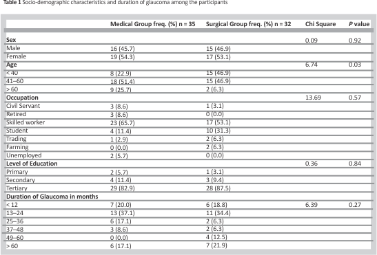
Thirty-five eyes of 35 patients on medical treatment for glaucoma and 32 eyes of 32 surgically treated patients were included in this analysis. The mean ages in the medical and surgical groups were 52.4 ± 14.8 years (range 19–82 years) and 39.3 ± 15.4 years (range 18–67 years), respectively (p value < 0.001). The majority of the participants (65.7% in the medical group and 53.1% in the surgical group) in both groups were skilled workers. Over 80% of participants in both groups had a tertiary level of education. Table 1 shows no statistically significant difference between the 2 groups in sex, occupation, or duration of glaucoma.
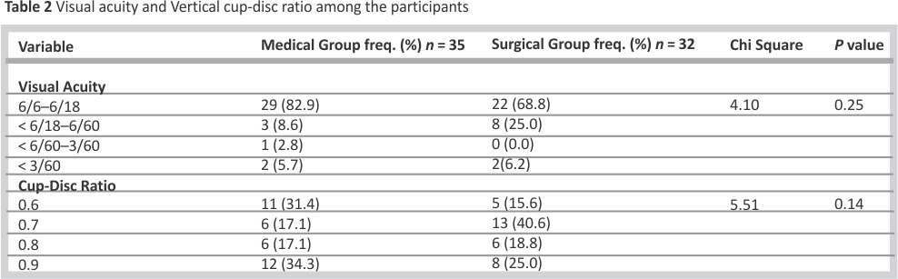
As depicted in Table 2, majority of the participants (82.9% in the medical group and 68.8% in the surgical group) had normal/mild visual impairment. There was no statistically significant difference between the 2 groups in visual acuity and vertical cup-disc ratio.
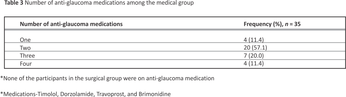
Over 85% of the participants in the medical group were on 2 or more medications, as shown in Table 3.
Also, across all measures of IOP fluctuations (peak IOP, trough IOP, IOP range, and change from baseline), the medical group exhibited significantly higher IOP values and more pronounced fluctuations compared to the surgical group (p value < 0.001) (Tables 4a and 4b). As highlighted in Table 4a, the Cohen’s d values demonstrate a large treatment effect across all evaluated parameters in favour of the surgical group with the largest effect observed for the peak IOP (d = 1.43) and trough IOP (d = 1.26).
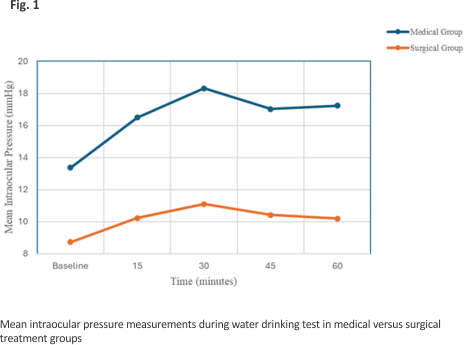
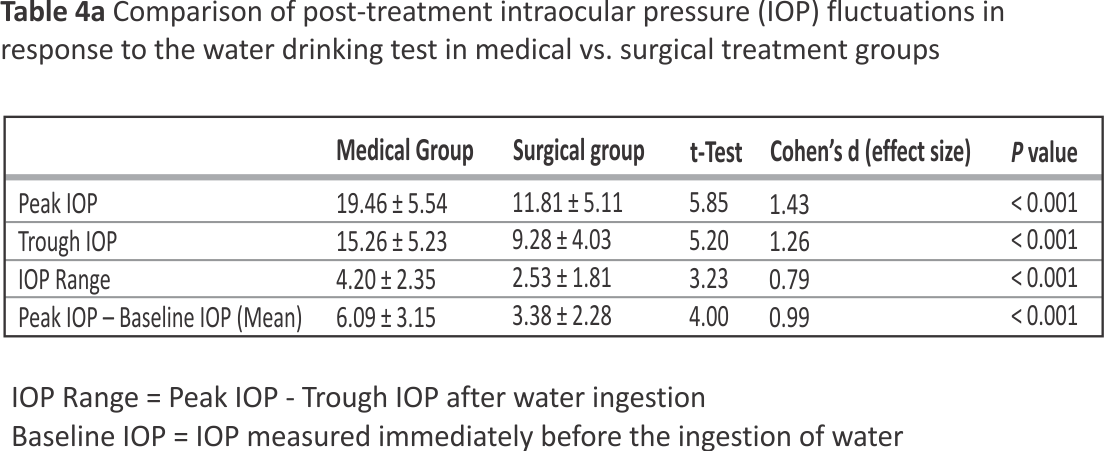
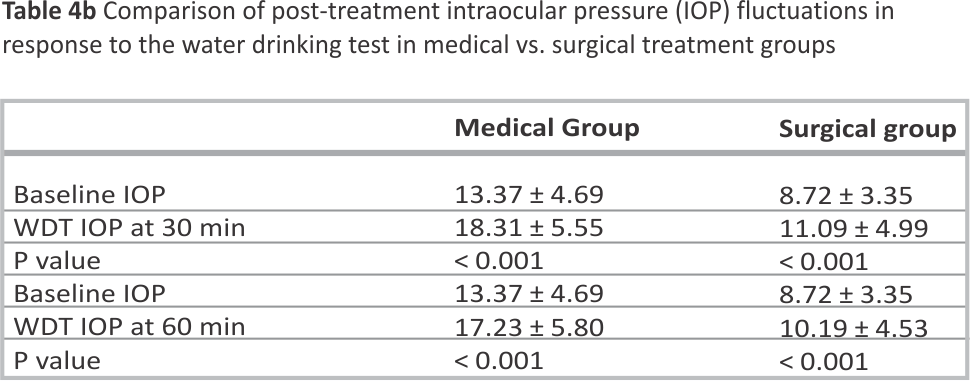
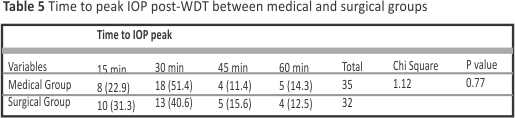

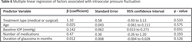
Patients in the surgical group were significantly younger than those in the medical group, ostensibly because younger patients were more likely to be offered surgical therapy as definitive management of their glaucoma, as has been documented by different studies 24. Multivariate analysis showed that although the surgical group was younger, the observed lower IOP fluctuation among surgically treated patients was not confounded by age or the other variables included in the model, except for baseline IOP. This study indicates that medically treated patients exhibited significantly higher mean IOP values and greater fluctuations across all measured parameters (peak IOP, trough IOP, IOP range, and change from baseline) compared to those in the surgical group (P values < 0.001), despite comparable baseline IOPs. These results show the superior efficacy of trabeculectomy in reducing IOP variability, which is recognized as a key factor in the progression of glaucoma 13. Fluctuations in IOP contribute to optic nerve damage even when average IOP appears well controlled,7 and the fact that the medical group experienced higher IOP fluctuations despite treatment suggests that medications may not be sufficient to maintain stable IOP over time. Trabeculectomy creates an alternative drainage pathway for aqueous humor drainage, offering superior and more stable IOP control, potentially reducing the risk of disease progression 10, 13. The greater IOP fluctuation seen in the medically treated group may be attributed to the limited ability of pharmacologic therapy to compensate for transient increases in aqueous inflow or episcleral venous pressure during the WDT. Most topical agents act by suppressing aqueous production or modestly enhancing uveoscleral outflow, mechanisms that rely on dynamic physiologic regulation and patient adherence. In contrast, trabeculectomy provides a passive alternative drainage route, allowing more consistent aqueous egress and dampening pressure spikes. This likely accounts for the smaller amplitude of IOP fluctuation and lower peaks observed in the surgical group. Similar findings have been reported by Muniesa et al.,19 who found that IOP-related fluctuations and IOP peaks were higher in eyes with medically treated glaucoma patients than in those treated surgically. A study by Medeiros et al. also using the WDT to assess IOP fluctuation observed that patients who had undergone trabeculectomy had less IOP fluctuations and significantly lower IOP peaks during the diurnal tension curve compared to medically treated glaucoma patients 20. The water-drinking effectively demonstrated IOP fluctuations in both groups, reflecting its role as an important tool for assessing true IOP control beyond static office measurements. Incorporating WDT into routine glaucoma management could help clinicians identify patients at higher risk for progression and adjust treatment plans accordingly.
Although this study focused on intraocular pressure fluctuations as a physiological outcome measure, it did not evaluate concurrent structural or functional progression parameters such as RNFL thickness or visual field change. However, prior studies—including the Advanced Glaucoma Intervention Study—have demonstrated that IOP fluctuation is an independent risk factor for glaucomatous progression, supporting its relevance as a surrogate marker of disease control. Future longitudinal studies correlating WDT-induced IOP fluctuations with visual and structural outcomes are warranted.
This study showed that there was a high dependence on multiple drug therapy to control intraocular pressure (IOP) as more than 85% of patients in the medical group were on two or more medications. Use of multiple antiglaucoma drugs (polypharmacy) could increase the risk of non-adherence to medications (due to side effects, cost, complex dosing regimens, and poor quality of life), further worsening IOP fluctuation and glaucoma progression 11. It may be logical to postulate that early surgical intervention may be beneficial, especially in patients with poor drug compliance associated high IOP fluctuation and glaucoma progression. This would reduce their long-term dependence on medications, which is associated with a huge financial burden in our resource limited environment.
The concept of interventional glaucoma, 25 an approach of managing glaucoma that goes beyond the traditional reactive approach where patients are only offered surgical therapy when medical treatments have failed, comes readily to the fore. This concept aims to circumvent the problems of adherence, local and systemic side effects by offering selective laser trabeculoplasty and minimally invasive glaucoma surgical procedures (MIGS) earlier in the course of the disease. The cost of procuring laser equipment and the cost of MIGS devices may make them beyond the reach of most patients in our setting, where most health expenditure is out-of-pocket. A study carried out among Nigerian glaucoma patients found that only 9.3% had access to health insurance, with the direct costs of routine glaucoma care taking up a significant percentage of the income of patients 26. Strengthening glaucoma services and performing trabeculectomies earlier may indeed play a major role in halting the inexorable progression of the disease towards blindness, with reduced long term dependence on medications. Studies have shown that primary trabeculectomy is more effective than primary medical treatment at lowering IOP and preventing disease progression even among patients with advanced disease, with similar quality of life outcomes 27.
New tools for early glaucoma screening like the new glaucoma screening calculators 28 that have been validated in patients of African descent may become critical for earlier detection and may enable intervention even earlier in the course of the disease. Earlier cataract surgery and microinvasive glaucoma surgery using the affordable sinskey hook goniotomy, in patients with concomitant cataract and glaucoma, are some cost-effective interventions that have been shown to be efficacious and may reduce medication burden among patients of African descent 29, 30.
Although the iCare rebound tonometer differs methodologically from the Goldmann applanation tonometer, previous studies 23 in the same population have confirmed their strong agreement, supporting its suitability for dynamic IOP monitoring during provocative testing.
In conclusion, the present study clearly shows that intraocular pressure (IOP) fluctuations and peak IOP levels are significantly higher in glaucoma patients managed with medications compared to those who underwent trabeculectomy. Surgical treatment, particularly trabeculectomy, offers more consistent and effective 24-hour IOP control, resulting in lower mean IOP, reduced peak IOP, and smaller IOP fluctuations. throughout the day. This suggests that trabeculectomy not only lowers IOP effectively but also stabilizes it over time, which is crucial in preventing glaucoma progression.
Limitations
The specific brands, type or number of antiglaucoma medications used by the patients in the medical group were not considered in the outcomes of the WDT, and stratification of the medical group by specific drug class was not performed, due to the limited sample size and extensive overlap from combination therapy. The only consideration was that the patients had achieved target pressures. Future studies with larger cohorts could evaluate the varying effects of the different drug classes on IOP fluctuations and WDT responses.
It is noteworthy that while the surgical group was assessed at least 6 months postoperatively, the medical group was assessed after at least 2 months of stable target IOP. This temporal mismatch reflects the different times to achieving stable IOP control for each treatment modality. The 6-month threshold ensured postoperative bleb stability, beyond the initial phase of IOP variability associated with transient inflammation, suture readjustment and bleb remodeling. The 2-month threshold for the medical group was consistent with literature defining pharmacological stable IOP control. Nevertheless, this difference is a potential limitation that merits consideration in the interpretation of the outcomes of this study.
Another limitation of this study is the absence of central corneal thickness (CCT) measurements, which can influence absolute IOP readings. However, since all measurements were performed using the same tonometer and under standardized conditions, relative comparisons of IOP fluctuation between groups remain valid. Future studies incorporating pachymetry would further strengthen the interpretation of IOP-related findings.
Data availability
The data supporting the findings of this study are available from the corresponding author, [TSO], upon reasonable request.
References
1. Gupta N, Weinreb RN. New definitions of glaucoma. Curr Opin Ophthalmol. 1997;8(2):38–41.
2. Wilson R, Richardson TM, Hertzmark E, Grant WM. Race as a risk factor for progressive glaucomatous damage. Ann Ophthalmol. 1985;17(10):653–9.
3. Martin MJ, Summer A, Gold EB, Diamond EL. Race and primary open-angle glaucoma. Am J Ophthalmol. 1985;99(4):383–7.
4. Coleman AL, Miglior S. Risk factors for glaucoma onset and progression. Surv Ophthalmol. 2008;53 (Suppl1):S3–10.
5. Heijl A, Leske MC, Bengtsson B, Hyman L, Bengtsson B, Hussein M, et al. Reduction of intraocular pressure and glaucoma progression: results from the early manifest glaucoma trial. Arch Ophthalmol. 2002;120(10):1268–79.
6. Saheb H, Ahmed IIK. Micro-invasive glaucoma surgery: current perspectives and future directions. Curr Opin Ophthalmol. 2012;23(2):96–104.
7. Kidd MN, O’Connor M. Progression of field loss after trabeculectomy: a five-year follow-up. Br J Ophthalmol.1985; 69(11):827–31.
8. Chauhan BC, Drance SM. The relationship between intraocular pressure and visual field progression in glaucoma. Graefes Arch Clin Exp Ophthalmol. 1992;230(6):521–6.
9. Kim SH, Lee EJ, Han JC, Sohn SW, Rhee T, Kee C. The effect of diurnal fluctuation in intraocular pressure on the evaluation of risk factors of progression in normal tension glaucoma. PLoS ONE. 2016; 11(10):e0164876.
10. Drance SM. Diurnal variation of intraocular pressure in treated glaucoma. Significance in patients with chronic simple glaucoma. Arch Ophthalmol. 1963;70:302–11.
11. Malihi M, Sit AJ. Effect of head and body position on intraocular pressure. Ophthalmology. 2012;119(5):987 –91.
12. Mosaed S, Liu JH, Weinreb RN. Correlation between office and peak nocturnal intraocular pressures in healthy subjects and glaucoma patients. Am J Ophthalmol. 2005;139(2):320–4.
13. Caprioli J, Coleman AL. Intraocular pressure fluctuation a risk factor for visual field progression at low intraocular pressures in the advanced glaucoma intervention study. Ophthalmology. 2008;115(7):1123– e11293.
14. Clement CI, Bhartiya S, Shaarawy T. New perspectives on target intraocular pressure. Surv Ophthalmol.2014;59 (6):615–26.
15. Susanna RJ, Clement C, Goldberg I, Hatanaka M. Applications of the water drinking test in glaucoma management. Clin Exp Ophthalmol. 2017;45(6): 625– 31.
16. Kumar RS, de Guzman MH, Ong PY, Goldberg I. Does peak intraocular pressure measured by water drinking test reflect peak circadian levels? A pilot study. Clin Exp Ophthalmol. 2008;36(4):312–5.
17. Wilensky JT, Zeimer RC, Gieser DK, Kaplan BH. The effects of glaucoma filtering surgery on the variability of diurnal intraocular pressure. Trans Am Ophthalmol Soc. 1994;92:373–7.
18. Wasielica-Poslednik J, Schmeisser J, Hoffmann EM, Weyer-Elberich V, Bell K, Lorenz K, et al. Fluctuation of intraocular pressure in glaucoma patients before and after trabeculectomy with mitomycin C. PLoS ONE. 2017;12(10): e0185246.
19. Muniesa M, Ezpeleta J, Benítez I. Fluctuations of the intraocular pressure in medically versus surgically treated glaucoma patients by a contact lens sensor. Am J Ophthalmol. 2019;207: 429–30.
20. Medeiros FA, Pinheiro A, Moura FC, Leal BC, Susanna Jr. R. Intraocular pressure fluctuations in medical versus surgically treated glaucomatous patients. J Ocul Pharmacol Ther. 2002;18(6):489–98.
21. Damji KF, Behki R, Wang L. Canadian perspectives in glaucoma management: setting target intraocular pressure range. Can J Ophthalmol. 2003;38 (3):189– 97.
22. Dhingra S, Khaw PT. The moorfields safer surgery system. Middle East Afr J Ophthalmol. 2009;16(3):112–5.
23. Ademola-Popoola DS, Odi AF, Akande TM. Comparison of IOP readings using rebound iCare tonometer and Perkins applanation tonometer in an African population. J West Afr Coll Surg. 2014;4(1):17–30.
24. Un Y, Imamoglu S, Alpogan O, Bolac R, Yildiz MB. Surgical approaches to juvenile open-angle glaucoma. Eur Eye Res. 2024;4(1):42–50.
25. Teo AWJ, Angmo D, Wong TTL, Nongpiur ME. Interventional glaucoma: rethinking glaucoma treatment paradigms in the Asian context. Indian J Ophthalmol. 2025;73(2):S207–213.
26. Bello SU, Eze UA, Umar MM, Abdull M, Babalola OE. Economic burden of glaucoma in nigeria: estimating the direct health care cost in a tertiary eye clinic. Nigerian J Ophthalmol. 2023;31(1):25–31.
27. King AJ, Hudson J, Azuara-Blanco A, Burr J, Kernohan A, Homer T, et al. Evaluating primary treatment for people with advanced glaucoma: five-year results of the treatment of advanced glaucoma study. Ophthalmology. 2024;131(7):759–70.
28. Grimes K, Madu CT, Carrington C, Laroche D. Validation of a novel Low-Cost glaucoma risk calculator for Community-Based screening in High-Risk populations. Clin Ophthalmol. 2025;19:357–69.
29. Calderon I, Laroche D. Affordable sinskey hook goniotomy and cataract surgery in black and afro-latino patients diagnosed with glaucoma: retrospective real-world one-year results. J Clin Med. 2025;14(10):3266. https://doi.org/10. 3390/jcm14103266. PMID: 4042926 2.
30. Zaifar A, Pratomo TG, Suryono AN. Comparison between MIGS with trabeculectomy in the management of open-angle glaucoma with cataract: A systematic review. Indian J Ophthalmol. 2024;72(Suppl 3):S345–53.
Acknowledgements
No acknowledgements were necessary.
Funding
The authors received no funding for this work.
Author information
Authors and Affiliations
Department of Ophthalmology, University of Ilorin Teaching Hospital, Ilorin, Nigeria
Tokunbo Sarah Obajolowo, Ibrahim Abiodun Yusuf, Ambali Olamilekan Ambali, Biola Toibat Tota-Bolarinwa & Fatai Olasunkanmi Olatunji
Obafemi Awolowo University Teaching Hospitals Complex, Ile Ife, Nigeria
Azeezat Bola Aderounmu
Department of Ophthalmology, University of Ilorin, Ilorin, Nigeria
Fatai Olasunkanmi Olatunji
Contributions
T.S.O., I.A.Y. and F.O.O. conceptualized and designed the work, A.O.A., B.T.B., A.B.A. drafted the initial manuscript, T.S.O. and A.O.A. were responsible for acquiring the data; I.A.Y. and A.O.A. were responsible for the analysis, and interpretation of data; T.S.O., I.A.Y., B.T.B., A.B.A., F.O.O. substantively revised the work. All authors read and approved the final submitted version of the manuscript.
Corresponding author
Correspondence to:
Tokunbo Sarah Obajolowo.
Consent for publication
Not applicable.
Competing interests
The authors declare no competing interests.
Credits: Obajolowo, T., Yusuf, I., Ambali, A. et al. Effect of medical and surgical therapy on intraocular pressure fluctuations among glaucoma patients in the University of Ilorin Teaching Hospital: A comparative clinical study. BMC Ophthalmol 26, 36 (2026). https://doi.org/10.1186/ s12886-026-04636-9